This unit explores the ways in which animals are adapted for survival and respond to changes in their internal and external environments. The emphasis is on vertebrates, particularly mammals, and explores the relationship between structure and function.
Mammalian nutrition
Breakdown of food
The main food groups are:
• Carbohydrates
– energy foods
• Proteins
– growth, repair and replacement of tissues
• Fats
– energy foods fats store twice the energy per gram compared to carbohydrates
• Vitamins
– complex molecules that take part in vital chemical reactions in cells
• Minerals
– simple molecules that take part in vital chemical reactions in cells.
Simple structure of foods
• Carbohydrates
Made of chains of glucose molecules.
Elements present – Carbon, Hydrogen and Oxygen (C,H,O)
• Proteins
Chains of amino acids.
Elements present Carbon, Hydrogen and Oxygen, and Nitrogen (C,H,O,N)
• Fats
A “backbone” of glycerol with three side chains of fatty acids.
Elements present – Carbon, Hydrogen and Oxygen C, H, O
Food tests
Food
Test
Change if positive
Reducing sugar
Benedicts reagent
When boiled change is from blue to brick red
Protein
Biuret test
Blue to violet/purple
Starch
Iodine
Orange to blue-black
Fats
Filter paper
translucent spot (light shines through but you can’t see things through it)
The need for digestion.
Digestion involves the breakdown of insoluble food substances into soluble food substances to allow absorption into the blood stream through the small intestine.
Function of the alimentary canal and associated organs
The alimentary canal is a continuous tube that connects the mouth to the anus.
Its primary function is the digestion and absorption of food.
Move your mouse over the graphic to learn the names of the parts.
Mouth
Mechanical breakdown of foods
Salivary glands
produce saliva, a solution of amylase and mucus:
Amylase digests starch down into maltose
Mucus lubricates the mouth and food to aid swallowing
Oesophagus
Joins the mouth to the stomach.
Food travels down by peristalsis
A wave of muscular relaxation followed by a wave of muscular contraction (see below)
Liver
stores excess glucose as glycogen:
Glycogen is made up of branched chains of glucose molecules
Like starch it is insoluble and is easily converted back to glucose
is the site of deamination:
Excess amino acids from the digestion of protein are broken down into energy compounds and urea.
Each molecule of urea contains two nitrogen atoms.
Makes bile which emulsifies fats:
Emulsify means that the fat is broken up into tiny droplets which have a much larger surface area for the enzyme lipase to act on (see below)
Gall bladder
Stores bile, which was made in the liver
Pancreas
produces three enzymes used in the small intestine:
Lipase – fats into fatty acids and glycerol
Trypsin – polypeptides into peptides
Amylase – starch into maltose
These enzymes are released into the start of the small intestine just after the stomach.
Stomach
Food is churned in stomach by action of longitudinal and circular muscles
which denature and begins digestion of proteins
Mucus – secreting cells protect the stomach lining from acid and enzyme attack.
Enzyme – secreting cells
produce pepsin that digests proteins down into polypeptides.
Acid – secreting cells produce acid that denatures the protein and provides optimum pH for pepsin.
Small intestine
Completes digestion of food
Food is absorbed into blood
Folded to increase surface area for absorption
Covered with villi which greatly increase the surface area
Large intestine
Connects small intestine to the anus
intestine
Reabsorbs water to produce faeces
Reabsorbs salts that have diffused out of the blood
Rectum
Stores faeces
Anus
Faeces egested (ejected)
Peristalsis
The alimentary canal is covered with circular muscles.
Food moves through the alimentary canal by:
a wave of muscular relaxation
followed by a wave of muscular contraction
Emulsification
The enzyme lipase can only act on the surface of the fat:
Fats are broken up into tiny droplets.
The chemical nature of the fat is not changed
This allows more lipase molecules to act on them
Absorption:
The surface of the small intestine is deeply folded to provide a large surface area for absorbing food into the blood.
The folds are covered in hair-like villi to further increase the surface area.
A villus has an excellent blood supply allowing rapid diffusion.
The thin membrane means there is a short distance between the blood and the food improves diffusion.
A villus is about 1 mm long and the entire surface of the small intestine is covered with them.
Amino acids and sugars diffuse into the blood capillaries
Amino acids and maltose enter these capillaries.
Products of fat digestion diffuse into the lacteal.
This is a finger like extension of the lymphatic system in the middle of the villus
Products of fat digestion enter the lacteal and are transported to the blood supply elsewhere.
Fate of food
after digestion and absorption:
Energy foods
carried in the blood to the cells
Sugars
starch
glycogen
other carbohydrates.
Fats
Excess energy food is stored first as glycogen in the liver then as fat around the body.
Proteins
For growth
For repair of damaged tissues
For replacement of worn out materials
excess protein is deaminated in the liver into energy carbohydrates and urea.
Control of the internal environment
The mammalian kidney
The kidney is the main organ for regulating water content in mammals (osmoregulation).
Water enters the body by:
Drinking
Food (there is some water in nearly all foods – e.g. oranges)
Metabolic Water (from respiration and other chemical reactions)
Water leaves the body by:
Urine
Sweat
Breathing
Faeces
Of these only urine production can be controlled to regulate the fluid in the body.
Urea
Urea is the waste product from breakdown of protein in the liver:
This process of breaking down protein is called deamination (see liver).
Urea contains nitrogen (and is therefore called nitrogenous waste)
Urea is transported to the kidneys in the blood
The kidneys remove the urea and dilute it with water to form urine.
Urine production
Move your mouse over the graphic to learn the names of the parts.
The functional unit of the kidney is the nephron made up of:
An area where the blood is filtered:
Blood is filtered out of the glomerulus (a bundle of capillaries within the Bowman’s capsule) into the Bowman’s capsule.
The Bowman’s capsule collects the filtrate.
Water, salts, sugars and urea pass into the kidney tubule
In the tubules useful dissolved materials are reabsorbed into the blood capillaries surrounding it.
In the loop of Henle more or less water is reabsorbed depending on ADH levels (see below).
Salts and sugars are reabsorbed
Urine is collected in the collecting duct to be passed to the bladder.
Negative feedback control by ADH
Negative feedback is a system used in many parts of the body where a steady state must be maintained.
ADH (antidiuretic hormone) affects the Loop of Henle controlling the volume of water in the urine:
Too low a water concentration in the blood:
Osmoreceptors in the hypothalamus are stimulated by a decrease in water concentration in the blood.
These trigger an increase in the release of antidiuretic hormone (ADH) from the pituitary.
The ADH enters the blood and is carried to the nephrons.
ADH increases the permeability of the kidney tubules and collecting duct:
More water is reabsorbed into the blood stream.
Little concentrated urine is produced.
Another action triggers the sensation of thirst.
Too high a water concentration in the blood:
Osmoreceptors in the hypothalamus are stimulated by a increase in water concentration in the blood.
These trigger an decrease in the release of antidiuretic hormone (ADH) from the pituitary.
The ADH enters the blood and is carried to the nephrons.
ADH decreases the permeability of the kidney tubules and collecting duct:
Less water is reabsorbed into the blood stream.
A larger volume of dilute urine is produced.
Another action reduces the sensation of thirst.
Osmoregulation in marine and freshwater bony fish
Marine bony fish have hypotonic tissues:
Water is lost continually through the gills by osmosis
Danger of dehydration
They are continually drinking salt water
Excess salt is excreted by specialised cells in the gills
Little, concentrated urine
Freshwater bony fish have hypertonic tissues:
Water is gained continually through the gills by osmosis
Danger of influx of water
Salt is lost through the gills
Salt is absorbed by specialised cells in the gills
Copious dilute urine
Circulation and gas exchange
The heart and blood vessels
Move your mouse over the second heart graphic to learn the names of the parts.
The heart is a muscular pump that moves blood around the body in a range of blood vessels.
There are four chambers in the heat:
The right atrium
blood received from the body, pumped into:
The right ventricle
blood is pumped to the lungs for oxygenation.
The left atrium
blood received from the lungs, pumped into:
The left ventricle
blood is pumped to the body.
The walls of the left ventricle are thicker than that of the right:
The right ventricle has a relatively short distance to pump blood to the lungs
The left ventricle has a relatively long distance to pump blood all round the rest of the body.
The AV valves (Atrio-ventricular) valves prevent blood flowing back into the atrium when the ventricles contract.
The AV valve in the right atrium is tricuspid, the AV valve in the left atrium is the bicuspid.
The semi lunar valves prevent blood flowing back into the ventricles when the ventricles relax.
The order of blood flow through the heart is:
Vena cava right atrium right ventricle
pulmonary artery lungs pulmonary vein
left atrium left ventricle aorta
Blood vessels
The heart obtains its blood supply from the coronary arteries.
Blockages of the coronary arteries lead to heart disease
Part of the hear muscles dies
This is commonly known as a hear attack
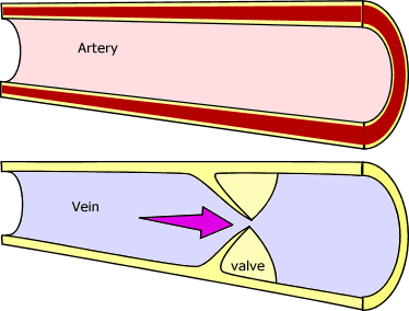
Blood leaves the heart in arteries
Arteries have thick, muscular walls to contain blood under high pressure.
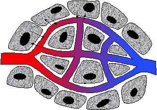
Blood flows through capillaries
Capillaries have very thin walls so that material can be exchanged with the tissues by diffusion
Blood returns to the heart in veins
Veins have thinner, less muscular walls than arteries
Blood is under lower pressure
Valves are present to prevent blood flowing backwards.
The pulse indicates that there is blood flowing in the artery below the point of testing.
The position and function of the following blood vessels should be known.
pulmonary artery and pulmonary vein (to and from the lungs)
aorta and vena cava (to and from the body)
hepatic artery, hepatic vein (to and from the liver)
mesenteric artery (to the small intestine)
hepatic portal vein (from the small intestine to the liver)
renal artery and renal vein (to and from the kidneys)
The coronary artery and vein (coloured red and blue respectively in the beating heart animation above) branches off the aorta and feeds blood to the heart itself.
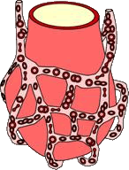
Lungs and capillary network
The lungs are the gas exchange structure in the mammal, here oxygen needed for cellular respiration is absorbed into the blood and CO2 waste from cellular respiration is passed out into the air.
Internal structure of lungs
Move your mouse over the graphic to learn the names of the parts.
The trachea carries air into and out of the lungs
The trachea branches into two bronchi (s. bronchus)
The bronchi branch into many bronchioles
The bronchioles end in millions of tiny air sacs – the alveoli (s. alveolus)
The lungs are excellent gas exchange structures because:
The many alveoli make a very large surface area
The inner surfaces of the alveoli are moist to allow oxygen to dissolve.
The walls of the alveoli are thin to allow rapid diffusion of dissolved gasses
The alveolus is surrounded by a capillary network for efficient collection of the oxygen and unloading of the CO2
Blood structure and function
Plasma
The plasma is a straw coloured liquid
It carries dissolved food, salts, hormones and some dissolved CO2
When CO2 dissolves it makes and acid solution, this limits the volume of CO2 that can be carried in the plasma.
Red Blood Cells (the smaller cells on the diagram)
Carry oxygen and some CO2
These are very small cells to fit into tiny capillaries
They contain the chemical haemoglobin that gives them their red colour.
Haemoglobin retains (binds to/collects) oxygen at high oxygen levels in the lungs to form oxyhaemoglobin
Haemoglobin releases oxygen at low oxygen levels in the tissues.
White Cells (the large cell with the large nucleus on the diagram)
There are many kinds of white cell but all are involved in the immune response (defence against germs and other disease causing organisms).
Of the many kinds we look at two types:
Lymphocytes produce chemical antibodies in respons
e to antigens on the surface of germs.
Antibodies are “Y” shaped molecules, the arms of the “Y” bind to the antigen sites on the membranes of the germ causing them to clump together.
An antigen is a chemical on the surface of a germ that is recognised as a target by a white cell.
There is a one antibody to match one antigen, this is produces by a particular variety of lymphocyte
In other words lymphocytes are specific – for every disease there is a strain of lymphocyte to produce the antibodies against it.
Macrophages detect the germs and destroy them by phagocytosis.
The membrane of the macrophage surrounds the germ.
The germ is sealed into a vesicle
The macrophage secretes digestive enzymes into the vesicle to digest the germ
Sensory mechanisms and processing of information
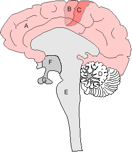
The brain
The cerebrum (A) is the folded surface of the brain, responsible for thought, conscious responses, memory, personality etc.
On either side of the sensory cleft on the cerebrum are:
The motor strip (B) dealing with movement
Sensory strip (C) dealing with touch.
The cerebellum (D) is responsible for balance and coordination.
The medulla (E) deals with heartbeat, breathing, peristalsis etc.
The hypothalamus (F) controls many regulatory functions such as body temperature and osmoregulation
The pituitary gland (G) produces a range of hormones including ADH.
The nervous system
The nervous system consists of the the CNS (Central Nervous System – brain and spinal cord) and the sensory neurones, motor neurones and senses.
The CNS sorts out information from the senses and sends messages to those muscles which can make the appropriate response.
Sensory neurones (nerves) carry information from the senses to the central nervous system.
Motor neurones (nerves) carry information from the central nervous system to the muscles.
Reflex action and the reflex arc.
When the body is in danger of damage the processing of the central nervous system is dangerously slow (around a quarter of a second or slower).
The reflex arc allows responses which are much faster and don’t involve processing by the CNS.
Critical information is detected by the receptors in the skin
Impulses travel up a sensory neurone (nerve) to the spinal cord
A relay fibre carries the impulse directly to an adjacent motor neurone
The impulse is carried along the motor neurone to appropriate muscles to carry out an appropriate response.
The response is rapid and protective
Temperature regulation as a negative feedback mechanism.
Negative feedback has been seen above regulating water balance in the body.
The body’s temperature is also regulated in this way:
Temperature receptors in the hypothalamus monitor body temperature.
If the body temperature rises:
Sweating is initiated
Blood vessels in the skin dilate (their diameter increases) to bring the warm blood close to the cooling sweat.
Erector muscles on the skin hairs relax, hairs lay flat, reducing the insulating air layer around the skin
If the body temperature drops:
Sweating is reduced
Blood vessels in the skin constrict (their diameter decreases) to keep the bulk of the blood deeper in the body.
Shivering occurs to provide heat energy from muscles’ respiration
Erector muscles on the skin hairs contract, hairs rise, increasing the insulating air layer around the skin
The result of this is to maintain a constant body temperature.