This topic serves as an introduction to some of the biological principles involved in the study of human movement and physical performance. It is intended that pupils will acquire knowledge and understanding of a range of processes associated with movement and of the relationships between physical activity and healthy living.
Movement
The purpose of this sub-topic is to provide a background to the primary
requirements of physical activity, i.e. support and movement. It is
intended to provide some understanding of how movement is achieved, of the
range of movement that is normally available and of the application of
these to physical activities.
The skeleton:
Provides a framework for support of the internal organs
Provides a system of levers on which to attach muscles and bring
about movement
Protects vital body organs such as:
Brain
Heart
Lungs
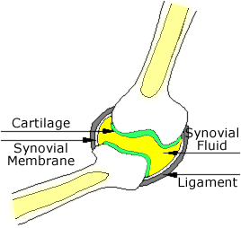
The Synovial Joints
Synovial joints are the joints which are able to move
They allow the bones to move against each other
The hinge joint:
Found at the elbow, knee and fingers
Permits movement in a single plane of movement (see textbook
for planes of movement)
The ball and socket joint
Found at the shoulder and between the hip and thigh
Permits movement in all three planes of movement.
Joints are held together by elastic ligaments
The structure of the synovial joint is shown.
The cartilage is smooth to reduce friction
The cartilage is spongy to absorb shocks
The synovial fluid is produced by the synovial
membrane and is oily to reduce friction in the join
Bone is made of:
Living cells which contain:
the hard mineral Calcium phosphate
flexible fibres of collagen
together these make a strong and durable material.
Muscles
attach to bones by inelastic tendons
tendons are inelastic so that the force of the muscle is applied fully and immediately to the bone –
that is they don’t stretch when the force
of the muscle acts on them.
Muscles work in opposing pairs
When the biceps in the arm contract the triceps
relax causing bending of the arm.
When the biceps in the arm relax the triceps contract
causing straightening of the arm.
Pairs of muscles are needed because the only active movement
of a muscle is to contract – to lengthen it must be stretched by
the action of an opposing muscle.
The purpose of this sub-topic is to give pupils the opportunity to
investigate systems of the body that provide muscles with the energy
resources that they require in producing movement. This involves
investigation of the structure of the respiratory and circulatory systems
and how these structures function in meeting the energy requirements of
muscles.
Energy
Our bodies gain energy through food
Our bodies expend energy in:
Movement
Heat
Chemical processes
Our bodied require to balance this energy:
If our energy intake is greater than our energy use:
Energy is stored as fat
There is a danger of obesity and its related diseases
If our energy intake is less than our energy output
Fat is converted to energy
We loose weight
There is a danger of starvation if the process continues for too
long.
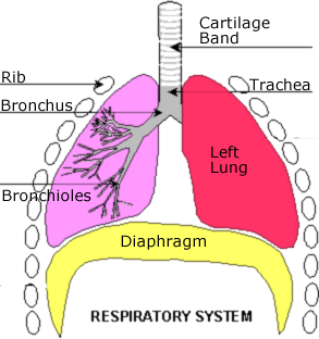
Breathing and the lungs
We obtain oxygen for respiration by breathing
During breathing we take oxygen into our blood During breathing oxygen is released from the blood into the exhaled air.
Structure:
The cartilage bands keep the windpipe open when our heads turn
and move
The trachea branches into two bronchi (s.
bronchus),
the bronchi branch into many bronchioles
The diaphragm is a flat sheet of muscle and connective tissue
that moves up and down to help breathing.
The ribs also are involved in breathing (see
below) as well as their protective role (see
above).
Each bronchiole ends in an air sac where gas exchange takes
place (see
below)
Breathing
To breathe in:
The muscles of the diaphragm contract causing the diaphragm
to move down
The intercostal muscles between the ribs contract causing
the ribs to move up and out
The volume of the chest increases and the pressure in the
lungs decreases
Air is drawn in.
To breathe out:
The muscles of the diaphragm relax causing the diaphragm to
move up
The intercostal muscles between the ribs relax causing the
ribs to move down and in
The volume of the chest decreases and the pressure in the
lungs increases
Air is forced out.
Cleaning
The tubes of the lungs (trachea, bronchi and bronchioles are lined
with:
Cilia (tiny, beating hair like structures)
Mucus producing glands
Germs, dust and dirt are trapped in the sticky mucus
The cilia carry the germs, dust and dirt out of the lungs
Gas Exchange
Takes place at the air sacs
These are microscopic sacs at the end of the bronchioles
Here oxygen diffuses into the blood
And carbon dioxide diffuses out of the blood
Air sacs are well adapted as gas exchange surfaces for the
following reasons:
The membranes of the airs sac and the capillaries are thin
(one cell thick)
The surface area is very large due to the very large numbers
of air sacs
There is an excellent blood supply due to the many
capillaries
The surface is moist to allow the oxygen to dissolve
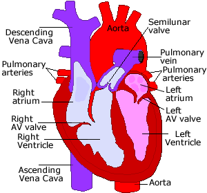
Circulatory system
The heart is a muscular pump which pumps blood around the body It
is made up of four chambers:
The left and right atria (s. atrium)
The atrium pumps blood into the ventricle
The left and right ventricles
The ventricles pump blood to the organs and tissues of the
body.
Blood follows a predictable path through the heart:
Blood enters the heart through the vena cava from the
body tissues and organs
It enters the right atrium which contracts to drive
the blood into:
The right ventricle, the ventricle contracts sending
the blood into:
The pulmonary artery, which carries the blood to the
lungs.
From the lungs the blood enters the pulmonary vein
which carries it to:
The left atrium which contracts to drive the blood into:
The left ventricle, the ventricle contracts sending the
blood into:
The aorta, which distributes the blood to the body’s
tissues and organs.
Within the heart four valves control the direction of blood flow:
From the atria into the ventricles
From the ventricles into the aorta and pulmonary artery
They prevent the blood flowing backwards during the relaxation of
the heart
The walls of the left ventricle are much thicker than the walls of
the right ventricle:
The right ventricle pumps blood a short distance through the lungs
The left ventricle pumps blood a long way through all the rest of
the body’s tissues and organs
The left ventricle must therefore be stronger and is therefore
thicker.
The muscles of the heart obtain their blood supply from the coronary
artery which is the first branch off the aorta just as it leaves the
heart.
The blood and its vessels
Blood vessels:
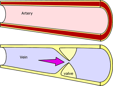
Blood travels away from the heart in arteries.
Arteries have thick, muscular walls to contain the blood
under pressure.
Blood flows through capillaries (see
below) where material is exchanged with the tissues.
Blood travels towards the heart in veins:
Veins have little muscle, and have valves to ensure that
the blood flows the correct way through the vessel.
The pulse indicates that blood is flowing in the artery under
the point of contact.
The blood:
The liquid part of the blood is the
plasma:
It is a clear, straw-coloured liquid. It carries
carbon dioxide, dissolved food and other soluble
chemicals
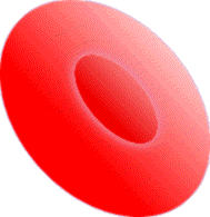
Within the blood are many red blood cells:
These are tiny cells adapted for moving
through very small blood vessels (capillaries) They carry
oxygen:
They are shaped like a ball with two dents top
and bottom.
Within the red blood cells is the chemical
haemoglobin.
In the lungs oxygen attaches to the
haemoglobin.
In the tissues the oxygen is detached from the
haemoglobin.
A red blood
cell
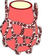
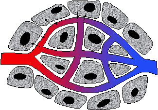
Gas and solute exchange
In the capillary network various chemicals are exchanged
between the cells and the blood by diffusion.
Oxygen and food enter the cells Carbon dioxide and waste
leave the cells
The capillary network has several features that make is well
adapted for exchange
The walls of the capillaries are thin (one cell thick)
for efficient diffusion
The network of capillaries gives a large surface area for
diffusion.
The network of capillaries gives an excellent blood supply
to the cells.
The purpose of this sub-topic is to allow pupils to measure some
physiological changes resulting from physical activity, and to use these
as indicators of level of performance and of fitness.
Fatigue
When a muscle or group of muscles is exercised continuously or
rapidly it gets fatigued.
When you exercise the muscle cells begin to respire faster to
provide more energy.
They need more food and oxygen to do this.
The heart rate and breathing increase to provide the muscle
cells with more food and oxygen.
There comes a point when heart, lungs and blood are not able to
supply enough oxygen for the muscle cells’ aerobic respiration
.
The muscle cells respire anaerobically.
When they respire this way they produce lactic acid.
Lactic acid build up in the muscles causes fatigue.
Anaerobic respiration:
sugar
lactic acid + Carbon dioxide.
Exercise
During exercise in an athlete the heart rate and breathing increase
less than in an untrained person.
Training improves the efficiency of the heart, lungs and
circulation
Recovery time is the time taken after exercise stops for
heart rate and breathing to return to normal.
During this time the excess lactic acid is changed back to
storage carbohydrate
Recovery time decreases as training improves the level of
fitness
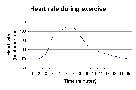
In the graph shown below, exercise begins at 2 minutes and stops
at 7 minutes.
Recovery time is from 7 minutes to 15 minutes.
Three factors can indicate the level of fitness of a person